V pátek 26. dubna 2024 úderem 22 hodiny začíná naše nová
a opravdu velká série soutěží o nejlepší webovou stránku !!
Proto neváhejte a začněte hned zítra soutěžit o lákavé ceny !!
a opravdu velká série soutěží o nejlepší webovou stránku !!
Proto neváhejte a začněte hned zítra soutěžit o lákavé ceny !!
Soubor:Diatoms-2004.png
Z Multimediaexpo.cz
Velikost tohoto náhledu je: 734 × 600 pixelů
Obrázek ve vyšším rozlišení (rozměr: 1 400 × 1 144 pixelů, velikost souboru: 951 kB, MIME typ: image/png)
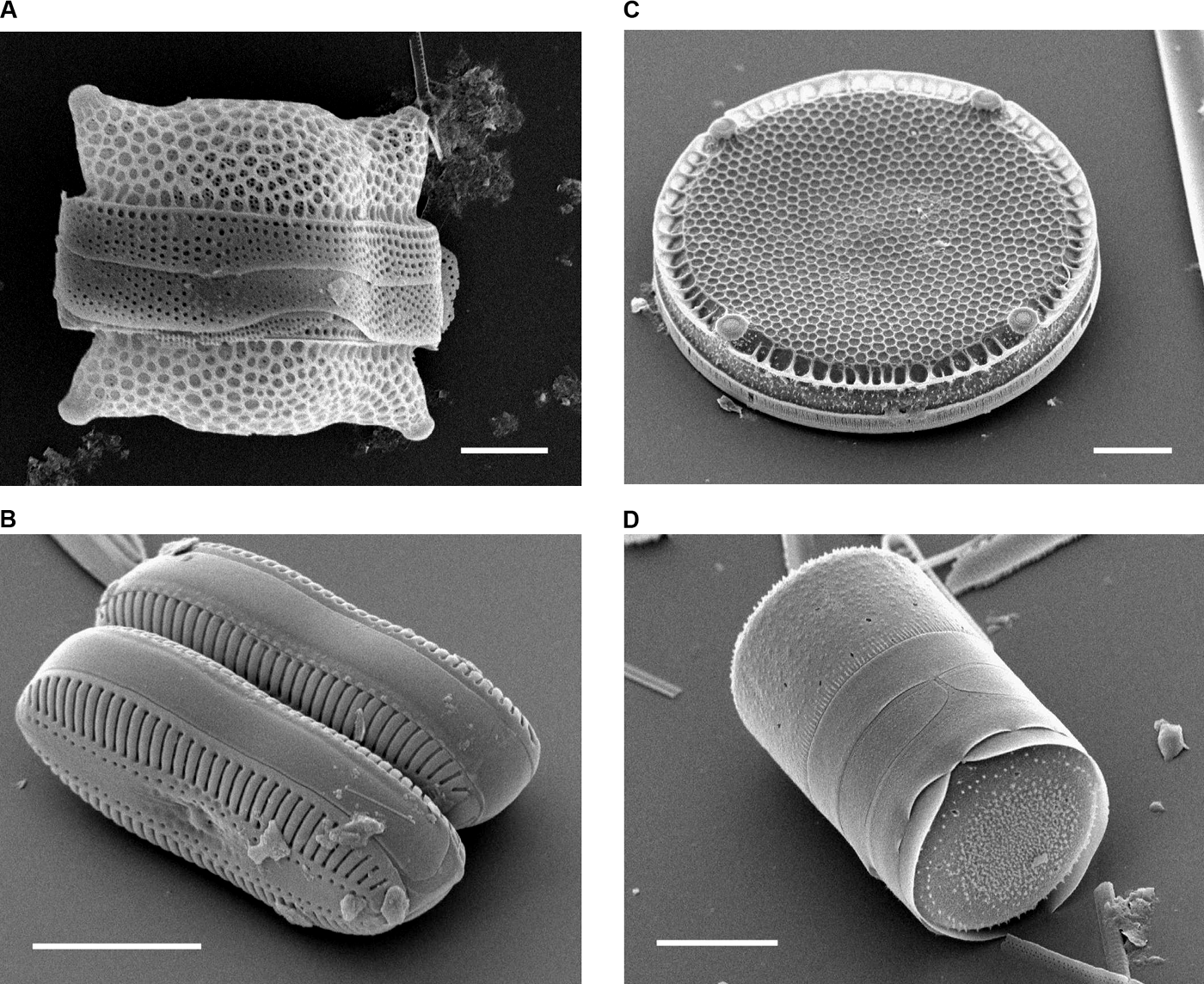
Fotografie + Description: Scanning Electron Micrographs of Diatoms.
- (A) Biddulphia reticulata. The whole shell or frustule of a centric diatom showing valves and girdle bands (size bar = 10 micrometres). (B) Diploneis sp. This picture shows two whole pennate diatom frustules in which raphes or slits, valves, and girdle bands can be seen (size bar = 10 micrometres). (C) Eupodiscus radiatus. View of a single valve of a centric diatom (size bar = 20 micrometres) (D) Melosira varians. The frustule of a centric diatom, showing both valves and some girdle bands (size bar = 10 micrometres).
- Date: Published: October 12, 2004
- Source: Bradbury J: Nature's Nanotechnologists: Unveiling the Secrets of Diatoms. PLoS Biol 2/10/2004: e306. doi:10.1371/journal.pbio.0020306
- Author: Images courtesy of Mary Ann Tiffany, San Diego State University.
+ pochází z Wikimedia Commons, kde má status – This file is licensed under the Creative Commons Attribution 2.5 Generic license. (CC BY 2.5)
Historie souboru
Kliknutím na datum a čas se zobrazí tehdejší verze souboru.
| Datum a čas | Náhled | Rozměry | Uživatel | Komentář | |
|---|---|---|---|---|---|
| současná | 2. 8. 2023, 06:29 | | 1 400×1 144 (951 kB) | Sysop (diskuse | příspěvky) | (Fotografie + ) |
- Editovat tento soubor v externím programu (Více informací najdete v nápovědě pro nastavení.)
Odkazy na soubor
Na soubor odkazuje tato stránka:


